Primary Cardiomyocytes
Study cardiac electrical activity, calcium handling, contraction and their functional relationships in the context of cell source, isolation and culture conditions.
Primary Cardiomyocyte Functional Assays

Covering primary cardiomyocytes, iPSC-derived cardiomyocytes and cardiac organoids, action potential recording, calcium imaging, voltage imaging, contraction and electrical-calcium-mechanical coupling support model characterization, pharmacological response studies and mechanistic research.
Confirm the compatibility of each model and assay with the sample conditions and project objectives.
Study cardiac electrical activity, calcium handling, contraction and their functional relationships in the context of cell source, isolation and culture conditions.
Primary Cardiomyocyte Functional AssaysCombine electrophysiology, Functional Imaging and contraction mechanics for multidimensional evaluation, from single cells to cell populations.
iPSC-Derived Cardiomyocyte Functional AssaysAnalyze functional phenotypes in cardiac organoids and cardiac-like tissues using electrical activity, calcium signals and contractile function measurements.
Cardiac Organoid and Cardiac Tissue-Like Model Assays| Functional Assay | Primary Cardiomyocytes | iPSC-Derived Cardiomyocytes | Cardiac Organoids |
|---|---|---|---|
| Patch-Clamp Action Potentials | Supported | Supported | Supported |
| Calcium Imaging | Supported | Supported | Supported |
| Voltage Imaging | Supported | Supported | Supported |
| Contractile Function | Supported | Supported | Supported |
| Electrical-Calcium-Mechanical Coupling | Supported | Supported | Supported |
Specific recording sites and assay combinations are determined by sample characteristics. Field potential, impedance and flexible-substrate mechanics assays require further adaptation to the culture configuration. Results from each assay are interpreted according to its measurement principle.
Directly record membrane potential changes to analyze action potential duration, waveform and rhythm-related parameters.
View serviceObserve calcium signal kinetics and functional changes following drug treatment to support calcium handling research.
View serviceUse voltage-sensitive probes to record electrical activity and analyze depolarization, repolarization and rhythm changes.
View serviceCombine optical motion analysis, compatible impedance-based beating measurements or flexible-substrate mechanics to evaluate contraction and relaxation.
View serviceCombine electrical activity, calcium handling and mechanical responses to identify the stages at which functional changes occur and the relationships between functional parameters.
View serviceUnder suitable culture conditions, observe population electrical activity, beating-related impedance and baseline state changes.
View serviceAnalyze calcium transients, rhythm characteristics and signal propagation in cardiac organoids and cardiac-like tissues to support functional evaluation and pharmacological response studies.
Explore Organoid Calcium Imaging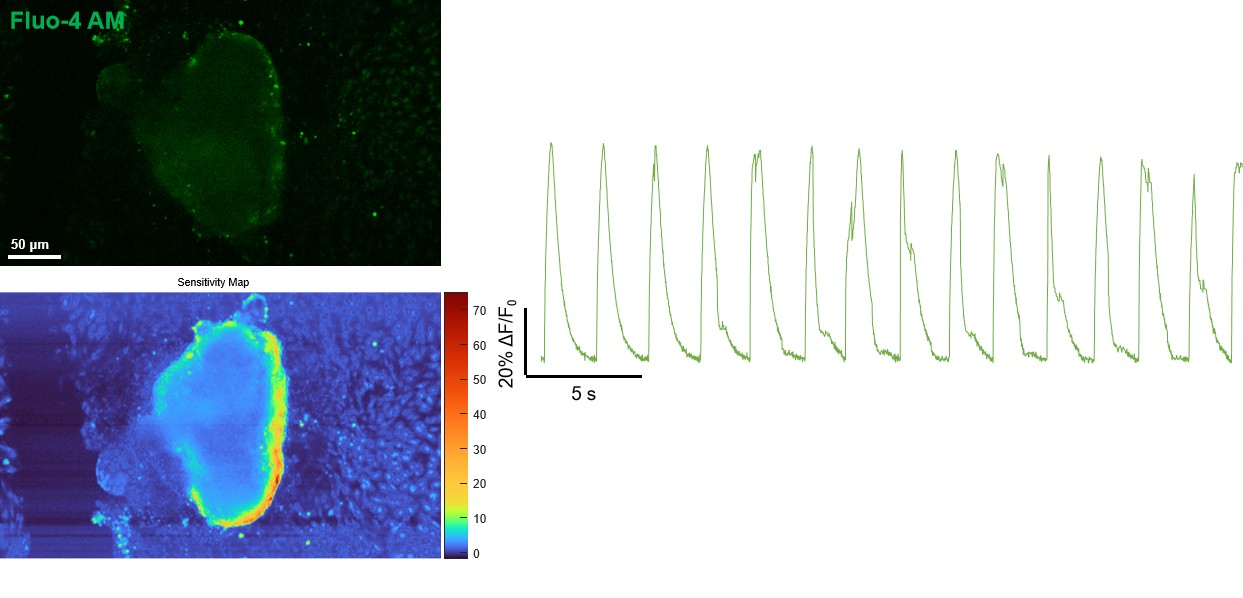
SparkMind (Nanjing) Technology Co., Ltd.
Rooms 1902–1904, 19th Floor, Building A, ZhongDan Eco Life Science Industrial Park
No. 3-1 Xinjinghu Road, Jiangbei New Area, Nanjing, Jiangsu Province, China
info@sparkmindcn.com
+86 25 5827 7019