
Assessing cardiac contractile function
Contraction is the mechanical output of cardiac electrical activity and calcium handling. We select optical motion analysis, beating-related impedance or flexible-substrate mechanics according to model structure and the research objective to investigate compound effects on contraction and relaxation.
Research models
Studies can use primary cardiomyocytes, iPSC-derived cardiomyocytes, cardiac organoids and cardiac-like tissues. Model origin, maturation in culture, tissue thickness and accessibility for measurement influence the experimental approach and interpretation.
Three complementary measurement approaches
| Approach | Key measurements | Interpretation |
|---|---|---|
| Optical motion analysis | Displacement or relative motion amplitude, contraction and relaxation velocities, durations and rhythm. | Suitable for cells or tissues whose motion can be tracked; motion amplitude itself is not contractile force. |
| Beating-related impedance | Beat-associated impedance changes, frequency, amplitude and rhythm stability. | Depends on cell-electrode contact, morphology and culture condition; impedance amplitude is not a direct force measurement. |
| Flexible-substrate mechanics | Substrate deformation, contraction and relaxation kinetics, and mechanical parameters derived using the appropriate calibration. | Force or stress is reported according to substrate properties, geometry and the calculation method, with explicit units and normalization procedures. |
Contractile function can be evaluated in primary cardiomyocytes, iPSC-derived cardiomyocytes and cardiac organoids. Suitability for impedance or flexible-substrate measurements must be confirmed for each sample based on adhesion, tissue anchoring and culture conditions.
Contraction and complementary functional signals
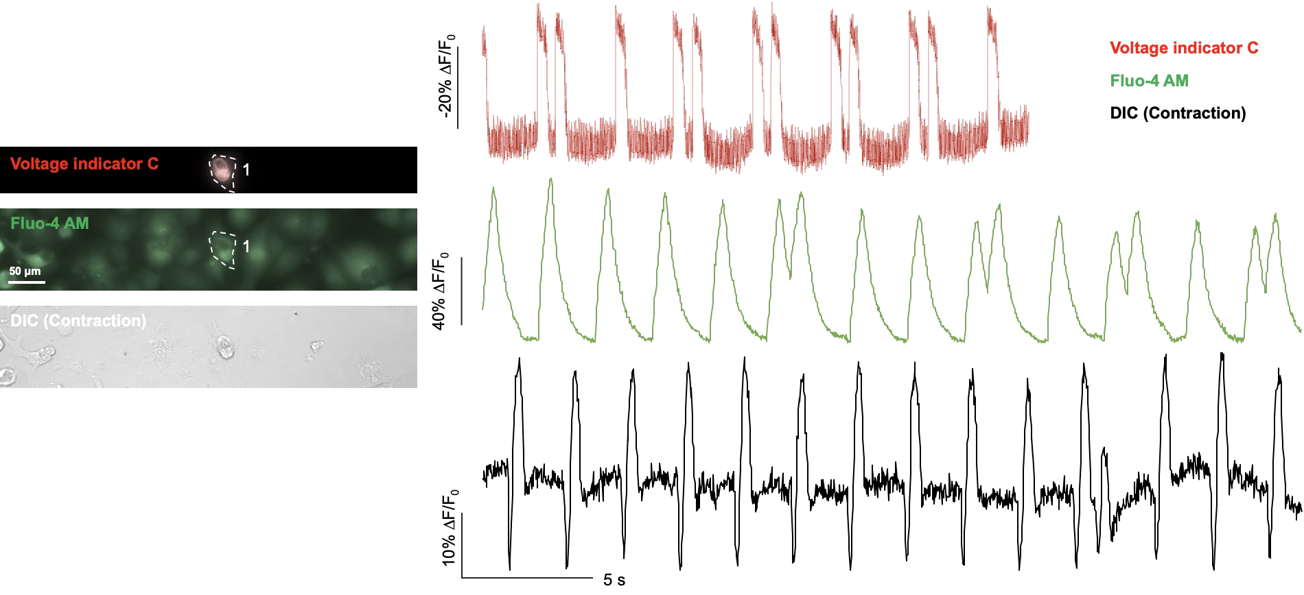
Combining electrical and calcium measurements with contractile assessment helps distinguish rhythm changes, altered calcium handling and abnormal contraction. The available measurement combination depends on model and assay compatibility; the example does not imply that all three signals can be recorded simultaneously in every model.
Project design and deliverables
The model, treatment concentrations, exposure times, control conditions and replicate design are agreed according to the study objectives. Results are analyzed alongside sample-condition and quality-control information.
- Documentation of the model, measurement approach, baseline condition and quality control.
- Contraction and relaxation parameters, concentration or time responses, and representative signals.
- Measurement units, calibration or normalization methods, and correlation analyses with other functional endpoints.