
Neuronal Voltage Imaging: Millisecond-Scale Signals and Circuit Mechanisms
Our neuroscience voltage imaging service emphasizes high temporal resolution and quantitative analysis of neuronal membrane-potential changes. Experimental and analytical support spans cell lines, primary neurons, brain organoids, brain slices and in vivo studies, with preparation-specific feasibility assessment. Studies can address neuronal interactions, network oscillations, dendritic and spine integration, and synaptic plasticity.
As a Functional Imaging approach, voltage imaging provides access to fast electrical activity and neuronal excitability beyond the temporal resolution of calcium-based measurements. Our existing workflows primarily use genetically encoded voltage indicators (GEVIs), with voltage-sensitive dyes considered where appropriate for the model and study design.
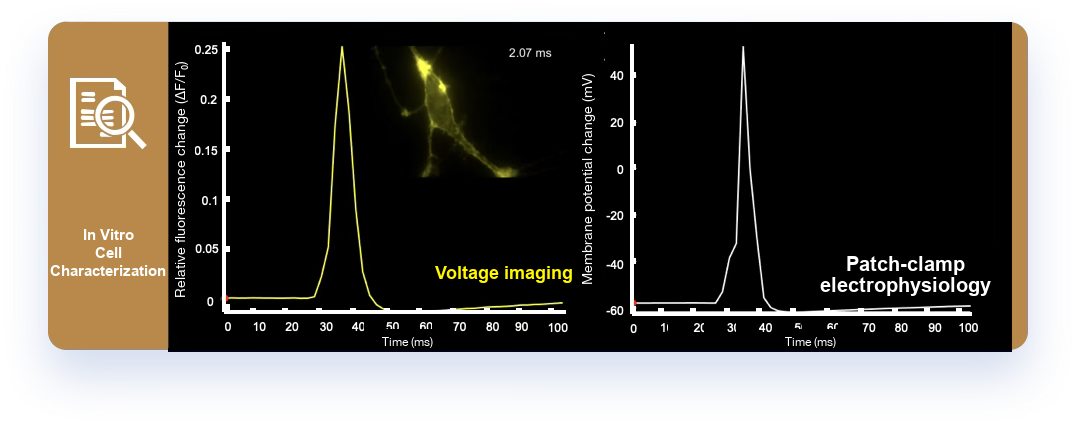
Core Service Capabilities
- Voltage imaging analysis: simultaneous recording and statistical analysis across multiple neurons to characterize interactions, synchrony, event-locked responses and network oscillations.
- Dendritic and spine voltage analysis: quantify depolarization and repolarization time courses, signal propagation and integration in selected dendritic or spine regions to investigate synaptic inputs and local processing, subject to spatial resolution and signal feasibility.
- All-optical stimulation and voltage imaging: coordinated stimulation-recording designs to test causal relationships and compare plasticity before and after induction, where optical stimulation and recording are compatible.
- In vitro, brain slice and in vivo voltage imaging: select the preparation for methodological validation, circuit-mechanism studies or behavior-related neural activity recording.
- Drug screening and mechanistic assessment: use voltage-associated endpoints for concentration-response and kinetic analysis in ion channel and neuromodulation research.
Model availability does not mean that every indicator, subcellular measurement or all-optical protocol is feasible in every preparation. Expression or labeling, optical access, motion and signal quality are assessed for the proposed endpoint.
Voltage Probes and Optical Electrophysiology
Voltage probes are molecular tools that track membrane-potential changes through voltage-dependent fluorescence. Unlike methods that measure intracellular calcium changes, they can more directly capture action potentials and subthreshold electrical signals, with millisecond-scale temporal resolution when the probe and acquisition system support it. This enables analysis closer to the kinetics of neuronal electrical signaling.
Two major probe classes are genetically encoded voltage indicators (GEVIs) and chemical voltage-sensitive dyes (VSDs). GEVIs support sustained expression and cell-type-specific labeling, making them useful for in vivo research. Voltage-sensitive dyes offer flexible and rapid labeling options for in vitro or slice experiments. Both can be paired with high-speed imaging to optically record neuronal electrical activity.
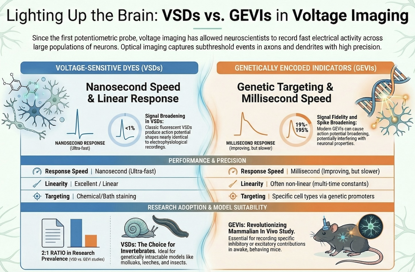
Optical electrophysiology uses light-based methods to record and modulate neuronal electrical activity. Voltage imaging enables simultaneous measurements from multiple neurons without contact from a conventional recording electrode, allowing analysis of firing patterns, oscillatory rhythms and intercellular interactions.
Voltage probes and optical electrophysiology help investigate neural circuit function, synaptic integration and behavior-related activity. Their temporal resolution and spatial coverage provide complementary ways to study how the brain processes information.
GEVI Workflows and an All-Optical Literature Example
GEVI-based workflows allow repeated optical monitoring of membrane-potential changes across neuronal populations, enabling analysis of action potential firing, subthreshold events and excitability modulation under different experimental conditions.
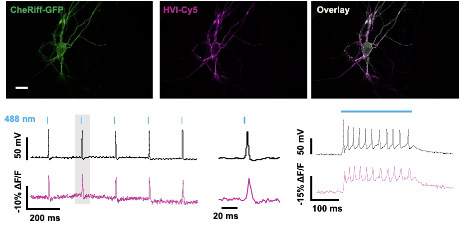
This literature example illustrates an all-optical approach; it is not a guarantee that the same probe and stimulation combination is suitable for every model.
Supported Cellular and Slice Models
- Heterologous cell systems expressing ion channels or receptors.
- Primary rodent neuronal cultures.
- Human iPSC-derived neuronal models.
- Acute rodent brain slice preparations.
- Rodent DRG sensory neuron cultures.
Brain organoid and in vivo study designs are also within the service scope described above, with model-specific feasibility assessment.
Functional Endpoints and Applications
- Spike frequency and firing patterns.
- Voltage-associated signal amplitude and kinetic changes.
- Subthreshold depolarization and excitability profiling.
- Comparative assessment of compound-induced effects.
- Exploratory functional phenotyping in disease-relevant models.
Network and Subcellular Questions
Voltage imaging extends single-cell observations to activity patterns across multiple neurons. Depending on the preparation, indicator expression and optical resolution, studies may examine firing synchrony, regional activation and voltage dynamics in selected dendritic regions.
Small dendritic and spine regions require feasibility assessment because signal strength, spatial resolution and motion can limit interpretation. Optogenetic stimulation can be combined with voltage recording in suitable all-optical experiments after checking spectral compatibility and stimulation artifacts. These capabilities do not imply universal all-optical compatibility across all brain slice or in vivo conditions.
Indicator Choice, Quality Control and Interpretation
GEVI workflows are selected according to expression requirements and response kinetics. Voltage-sensitive dyes can provide a complementary approach when suitable for the model and study design.
Fluorescence recordings generally report relative voltage-associated changes rather than calibrated absolute membrane potentials. Sampling rate, indicator kinetics and signal-to-noise ratio constrain spike and subthreshold measurements. Quality control addresses bleaching, motion, baseline drift and optical artifacts; analysis settings and exclusions accompany traces and summary results.