
Cardiac Voltage Imaging
Voltage-sensitive probes convert changes in membrane potential into optical signals, allowing electrical activity to be observed in suitable cells or tissue regions. Complementing single-point electrophysiological recordings, voltage imaging supports regional comparisons and studies of spatial electrical activity.
We provide cardiac voltage imaging for cardiovascular research and early discovery, with standardized acquisition, interpretable functional parameters and clear data visualization. Applications include exploratory evaluation of action potential behavior and rhythm stability, comparison of compound-induced membrane-potential changes, analysis of propagation in suitable multicellular preparations, and mechanistic studies of cardiac excitability and repolarization.
Imaging Approaches
- Voltage-sensitive dyes for recording rapid membrane-potential changes.
- Genetically encoded voltage indicators (GEVIs), where suitable for the model and project.
- Optional pacing or stimulation formats selected according to the study objectives.
Supported Research Models
The service covers primary cardiomyocytes, iPSC-derived cardiomyocytes, and cardiac organoids or cardiac tissue-like preparations. Model source, culture maturation, tissue thickness and optical access influence the approach and interpretation. Preparations can include suitable neonatal or adult primary rodent cardiomyocytes and human iPSC-derived cardiomyocytes in study-appropriate culture formats. Organoid or microtissue measurements require adequate staining or indicator expression and optical access.
Sample preparation, maturation, spontaneous or paced rhythm, temperature and exposure conditions are considered in comparisons. Recording from an accessible organoid region does not establish the electrical behavior of the entire three-dimensional tissue.
Key Evaluation Parameters
| Evaluation area | Analysis scope |
|---|---|
| Optical action potentials | Analyze waveform morphology and action potential duration when signal quality is suitable. |
| Rhythm characteristics | Assess beat frequency, beat-to-beat consistency and patterns of abnormal electrical activity. |
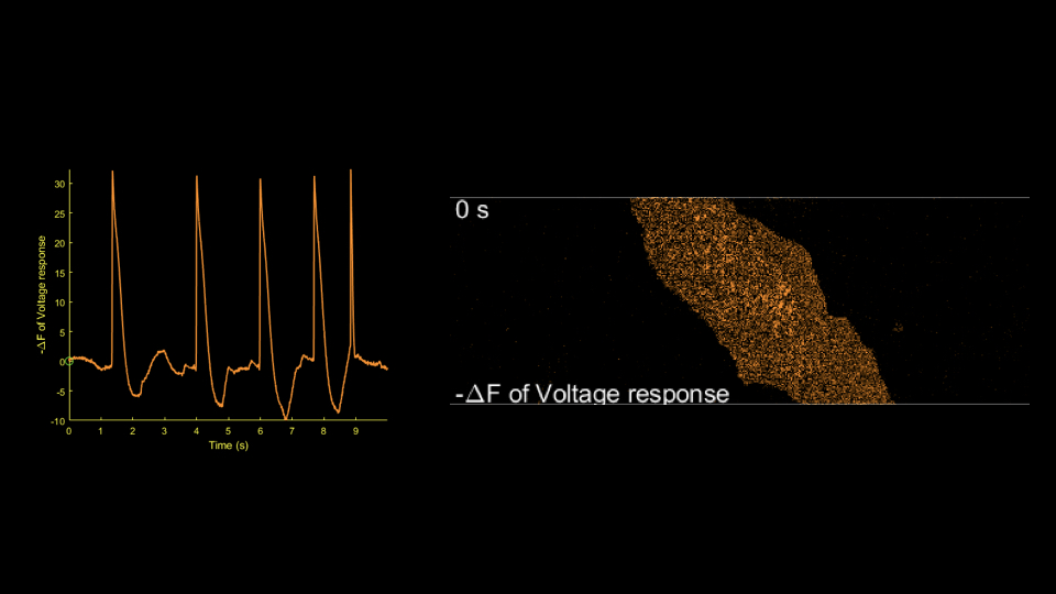
| Regional electrical activity | Compare activation timing, synchrony and propagation characteristics across resolvable regions. |
| Functional relationships | Compare changes in electrical activity with calcium transients and contractile responses. |
Optical signals generally reflect relative membrane-potential changes. Absolute membrane potential, extracellular field potentials and optical action potentials must be interpreted as distinct measurements. Analysis of rapid processes such as the upstroke is limited by acquisition speed, indicator response and signal-to-noise ratio.
Where supported, analysis includes optical action potential duration at defined repolarization levels, waveform changes and rhythm irregularity. Abnormal events require trace review, and propagation analysis requires sufficient spatial and temporal sampling and signal quality.
Electrical Activity and Other Functional Signals
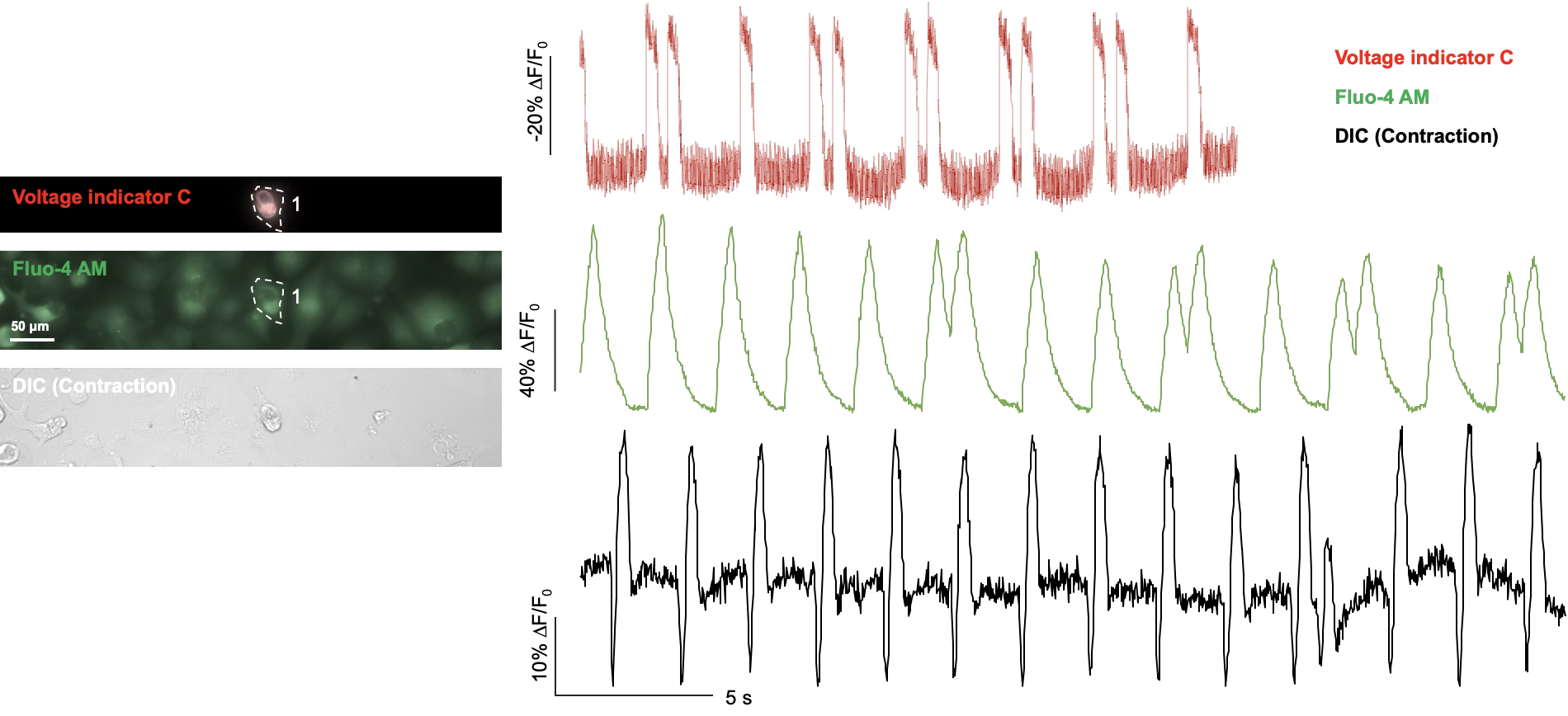
Cardiac Voltage Imaging Example
Voltage results can be compared with calcium and contraction measurements to investigate the relationship between excitation and mechanical activity. Compatibility is evaluated for each model; combined analysis does not imply that all signals are always acquired simultaneously or that motion-derived contraction is a direct force measurement.
Study Design, Quality Control and Deliverables
The model, treatment concentrations, exposure times, controls and replication are agreed according to the research objectives. Results are analyzed alongside sample condition and quality-control information. Analysis accounts for motion, photobleaching, background fluorescence, baseline drift and pacing or illumination artifacts.
- Description of the probe, acquisition conditions, regions of interest and signal quality.
- Optical electrical-activity traces, agreed parameters, concentration- or time-response results and quantitative summaries.
- Explanation of the effects of photobleaching, motion and acquisition conditions on interpretation.
- Methods, ROI definitions, representative traces and documented exclusions accompanying the results.