
Calcium imaging in cardiac organoids
Fluorescent calcium indicators reveal changes in calcium signals in cardiac organoids and cardiac-like tissues. Analysis of local transients, consistency of regional activity and functional responses to drug treatment supports characterization of three-dimensional cardiac models and pharmacological research.
Measurements and analysis
| Area | Analysis |
|---|---|
| Temporal kinetics | Calcium transient amplitude, rise and decay, frequency, and beat-to-beat variation. |
| Regional comparisons | Compare changes in signal intensity, activation timing and activity consistency across regions of interest. |
| Spatial propagation | Analyze propagation characteristics where spatial resolution, acquisition speed and signal quality are sufficient. |
| Drug responses | Compare calcium-signal phenotypes before and after treatment, or across concentrations and exposure times. |
Fluorescence signals are affected by tissue thickness, probe distribution and motion. Conclusions apply to the regions that can actually be observed and cannot be directly generalized to a uniform state throughout the entire three-dimensional tissue.
Calcium imaging example
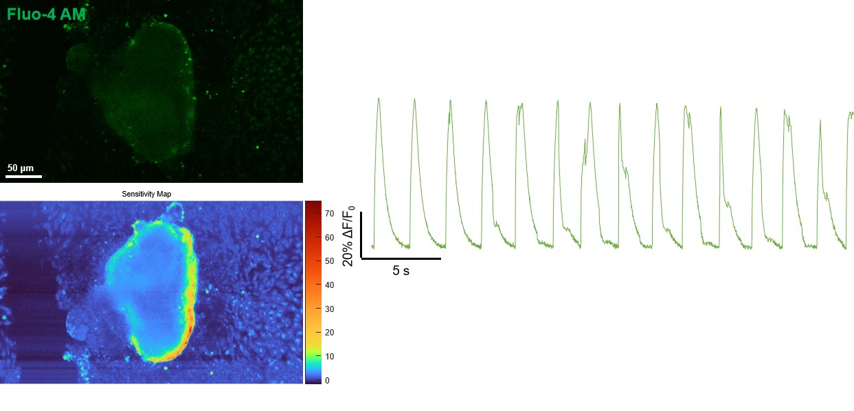
Linking calcium signals to broader functional assessment
Calcium imaging is one component of cardiac organoid functional assessment. Combining it with patch-clamp action potential recordings, voltage imaging, contraction measurements and electrical-calcium-contraction coupling studies can clarify the relationship between altered calcium handling and other functional processes. Assay combinations are selected for model compatibility; integrated analysis does not imply simultaneous acquisition of all modalities in every model.
Project design and deliverables
The model, treatment concentrations, exposure times, control conditions and replicate design are agreed according to the study objectives. Results are analyzed alongside sample-condition and quality-control information.
- Description of sample condition, probes, acquisition settings and regions of interest.
- Representative images and calcium transient traces, agreed functional parameters and regional comparisons.
- Correlation analysis with other functional measurements and an explanation of sampling limitations.