
Voltage Imaging and Membrane Potential Dynamics
Changes in membrane potential are fundamental to neuronal information encoding and cardiomyocyte electromechanical activity. Action potentials and subthreshold voltage fluctuations shape excitability, signaling efficiency and coordinated network activity. Voltage imaging uses voltage-sensitive dyes or genetically encoded voltage indicators (GEVIs) to convert millisecond-scale membrane potential changes into optical signals, enabling simultaneous monitoring of fast electrical activity at multiple sites without recording electrodes entering each cell. Our optical imaging platform supports both conventional voltage-sensitive dyes and GEVI-based approaches, providing high-temporal-resolution functional measurements for neuroscience, cardiac safety evaluation and ion channel research.
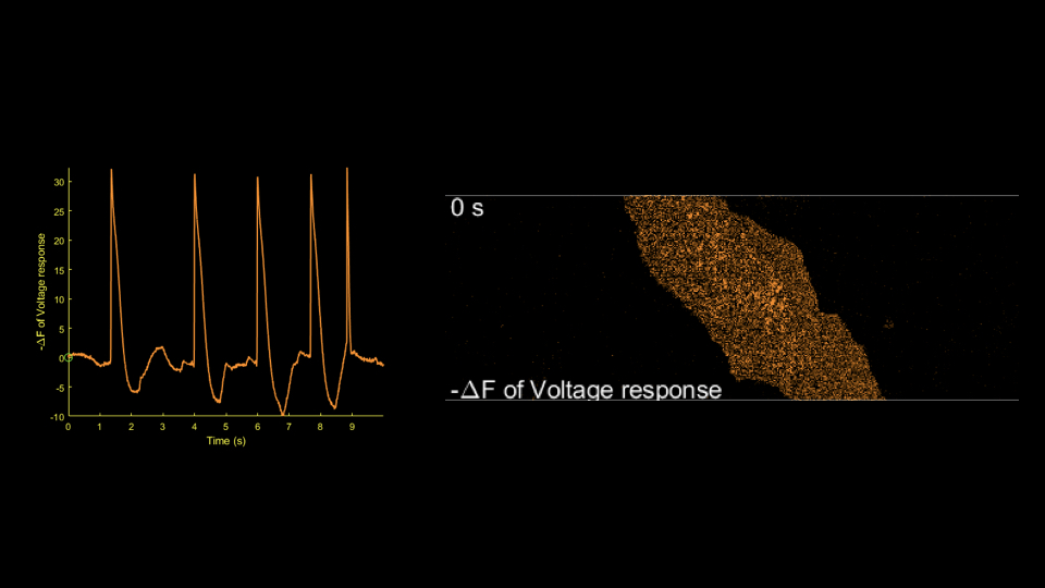
The full video is available on request.
Principles and Platform Capabilities
- Voltage-sensitive dye imaging: Small-molecule fluorescent dyes sensitive to membrane potential track action potentials and fast depolarization or repolarization with high temporal resolution.
- GEVI imaging: Genetically expressed voltage-sensitive fluorescent proteins support longitudinal, cell-type-specific membrane potential recordings for chronic experiments and network studies.
- Millisecond temporal resolution: Acquisition is optimized for fast electrical signals to capture individual action potentials, changes in firing frequency and subthreshold activity.
- Simultaneous multicellular recording: Single cells and cell populations can be imaged to analyze neuronal network synchrony and propagation.
- Standardized, extensible workflows: Signal-to-noise ratio and reproducibility are controlled, with drug treatment, genetic manipulation or structural imaging incorporated to improve interpretation.
Supported Model Types
- Neuronal models: Cell systems expressing cloned ion channels; rodent primary neuronal cultures; human iPSC-derived neurons; and brain slices or neuronal network models with GEVI expression.
- Cardiac models: Primary cardiomyocytes and human iPSC-derived cardiomyocytes (iPSC-CMs) for assessing action potential duration, depolarization and repolarization kinetics, and conduction properties.
- Other excitable cells: Feasibility is assessed against project requirements.
Research Applications
- Neuroscience: Directly observe action potential firing patterns, firing frequency and network synchrony. Investigate immediate effects of compounds or genetic manipulation on neuronal excitability.
- Cardiac safety and efficacy: Quantify action potential duration (APD), abnormal repolarization and risks associated with conduction disturbances, providing electrophysiological evidence for cardiotoxicity assessment.
- Ion channel and receptor function: Use membrane potential as a direct functional endpoint to investigate modulation of voltage-gated sodium, potassium and calcium channels, supporting mechanistic studies and candidate compound screening.
- Complementarity with calcium imaging: Combine voltage measurements with calcium transient data to connect electrical signals, calcium signals and functional phenotypes.
Integrated Voltage Imaging and Quantitative Analysis
Our platform supports both conventional voltage-sensitive dyes and GEVI voltage imaging, balancing temporal resolution, stability and experimental flexibility. We emphasize data quality control and parameter-based analysis, tailoring acquisition and analysis to research objectives. Integration with calcium imaging and electrophysiology provides interpretable electrical-function measurements for research and drug development.