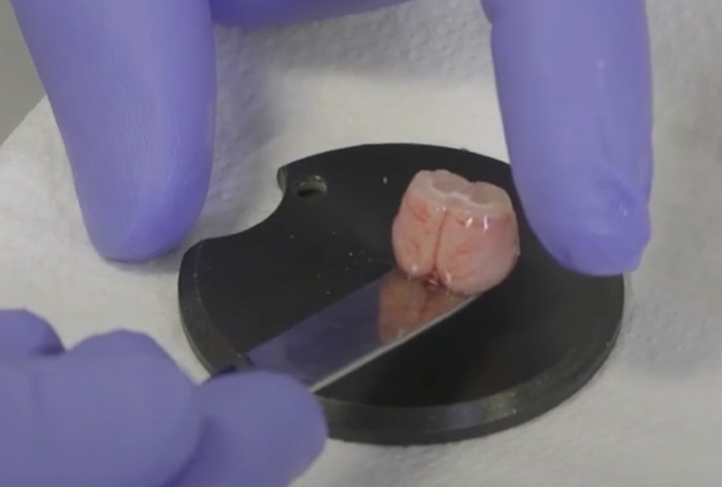
Acute Brain Slice Studies
Patch-clamp electrophysiology is a core technique in neuroscience and cellular physiology and a gold-standard method for measuring cellular electrical activity, particularly the electrical properties of individual neurons. Brain slice patch-clamp recording measures neurons in situ in freshly prepared tissue, combining single-cell resolution with preservation of local tissue architecture. It helps connect molecular mechanisms with circuit-level function. We aim to prepare healthy acute slices containing well-preserved, plump neurons suitable for patch-clamp recording.
Using acute brain slice models, we combine patch-clamp electrophysiology and calcium imaging to investigate passive and active membrane properties, synaptic transmission and plasticity, and firing activity in local neural circuits, supporting neuroscience research and drug discovery.
We perform electrophysiological recordings in acute rodent slices across a broad range of regions, including:
- Cortex, hippocampus, striatum, locus coeruleus, thalamus and amygdala
- Dorsal raphe, periaqueductal gray, cerebellum and spinal cord
- Substantia nigra, dorsal motor nucleus of the vagus and ventral tegmental area
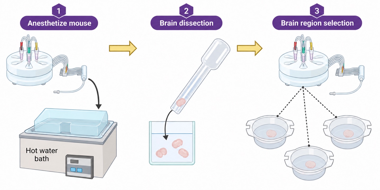
Patch Clamp: Passive Membrane Properties
- Input resistance
- Membrane capacitance
- Access resistance
- Resting membrane potential (RMP)
Patch Clamp: Active Membrane Properties
- Rheobase
- Action potential threshold
- Action potential amplitude
- Firing frequency
- Voltage-gated ion channels
- Ligand-gated ion channels
- G protein-coupled receptor (GPCR) modulation
Patch Clamp: Synaptic Transmission and Plasticity
- Evoked responses
- Miniature currents
- Spontaneous currents
Calcium Imaging
Monitor activity-associated calcium signals in small neuronal populations to investigate firing-related activity. Calcium signals are an indirect measure of electrical activity, not a direct membrane-voltage recording.
Applications
- Target functional validation: record ion channel- or receptor-mediated currents to assess target function following gene editing or drug treatment.
- Electrophysiological assessment of drug effects: evaluate modulation of neuronal excitability and synaptic transmission under near-physiological conditions.
- Disease-model mechanisms: characterize functional abnormalities in neurons from specific brain regions in animal disease models.
- Synaptic plasticity: induce and record long-term potentiation (LTP) and long-term depression (LTD) to investigate mechanisms relevant to learning and memory.
Brain Slice Patch-Clamp Recording Services
A typical workflow begins with acute slice preparation and recovery, followed by transfer to a recording chamber containing oxygenated artificial cerebrospinal fluid (ACSF). Target neurons are identified under a microscope; a glass electrode is positioned against the cell membrane using a micromanipulator to establish a high-resistance seal, after which neuronal electrical activity is recorded in real time. Detailed measurements from individual or small numbers of cells can help reduce reliance on large numbers of animals while supporting cellular mechanistic analysis.
Brain slice patch-clamp services cover mouse, rat, non-human primate and brain organoid slices, supporting functional investigation of multiple regions such as the hippocampus. Model, region, preparation and endpoint compatibility are established for each study; the listed measurements are not assumed to be feasible in every model.
- Ion channel function: record voltage- and ligand-gated currents, including sodium, potassium, calcium and chloride channel currents, as well as glutamate- and GABA-receptor-mediated currents.
- Synaptic function: record excitatory and inhibitory postsynaptic currents (EPSCs/IPSCs) to assess synaptic transmission and the effects of pharmacological intervention.
- Neuronal excitability: use current clamp to characterize action potential firing, thresholds and resistance, with optogenetic or chemogenetic stimulation where compatible with the preparation.
- Synaptic plasticity: induce and record LTP, LTD and paired-pulse ratio (PPR) measurements for studies of learning, memory and synaptic regulatory pathways.
Measurements and Study Design
Current-clamp recordings can characterize resting membrane potential, action potential threshold, firing frequency and responses to injected current. Voltage-clamp studies can assess selected ion channel currents and spontaneous or miniature synaptic currents; the recording configuration and pharmacological isolation are chosen for the endpoint.
- Specify the brain region, animal age, genotype or disease model, and target cell population before slice preparation.
- For synaptic studies, define stimulation sites, holding potentials and the intended EPSC, IPSC or plasticity measurements.
- Compare vehicle and treatment conditions using predefined recording windows and appropriate biological replication.
Recording Quality and Data Delivery
Slice viability, recovery conditions, recording temperature, seal quality and access resistance influence data interpretation. Acceptance criteria and exclusions are documented; unstable recordings are not treated as equivalent to stable recordings.
Deliverables can include representative traces, analyzed electrophysiological parameters, group summaries and a methods report describing preparation, recording conditions and analysis. A slice preserves local circuitry but does not reproduce the full connectivity or systemic environment of the intact brain.