
Customizable Histology Solutions
We process animal samples supplied by clients and can assist with sourcing specified laboratory mouse strains. Supported specimens include tissues from commonly used rodents and other species. We have particular experience in brain and spinal cord collection and sectioning, as well as sampling and analysis of liver, kidney, spleen, lung, muscle, intestine, skin and other organ systems. Sectioning plans can be customized for section orientation, thickness, collection interval, number of serial sections and mounting orientation.
Tissue samples are processed under controlled temperature and humidity in a contamination-controlled environment. Standardized equipment and established procedures support data comparability and traceability. We continually develop and validate tissue-marker and antibody combinations, with literature searches, marker selection, staining optimization and label validation tailored to different research questions. The platform maintains hundreds of validated antibodies and probes for varied labeling combinations and tissue-specific detection.
Multichannel fluorescence imaging is available. TSA staining kits can be used to label different antibodies together with DAPI nuclear staining for cellular localization and quantification. Automated acquisition balances speed and resolution and supports whole-slide scanning and high-resolution microscopy. Standardized image-analysis workflows provide rapid, objective and reproducible quantitative results. Analysis can also be customized to the marker features or regions relevant to your study.
Our experienced technical team performs sample processing and analysis in-house using standardized workflows, with appropriate cold-chain transport arrangements to protect sample integrity. We aim to maximize the information obtained from each specimen and provide high-quality, reproducible histology data to support publication and registration-related study requirements. Suitability for a specific submission must be assessed against that study's requirements; these are research services, not clinical diagnostic services.
Histopathology Service Portfolio
Tissue Processing
Professional tissue processing supports morphological and molecular integrity and specimen traceability. Fixation, dehydration, clearing and paraffin embedding produce high-quality tissue blocks for routine histology, immunohistochemistry (IHC), in situ hybridization (ISH) and other downstream experiments.

Routine Histology
High-quality hematoxylin and eosin (H&E) staining and a full range of special stains reveal tissue architecture for disease-model studies, drug toxicology assessment and morphological analysis.
Explore routine histology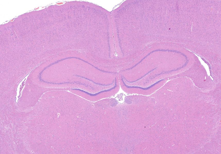
Molecular Pathology
Molecular pathology services include immunohistochemistry and immunofluorescence (IHC/IF), ISH including RNAscope®, and TUNEL apoptosis detection. These support gene and protein localization, expression assessment and apoptosis evaluation, combined with expert pathological interpretation and automated image analysis.

Spatial Biology
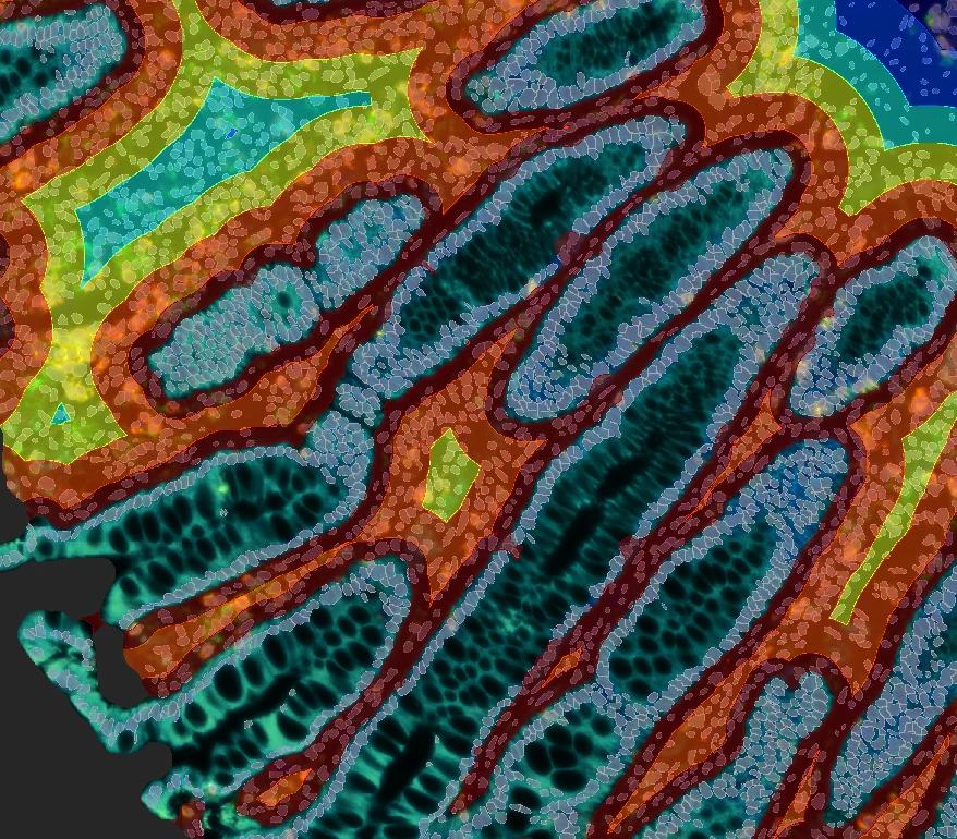
Multiplex-labeling-based spatial biology services support high-throughput, multitarget detection in FFPE and frozen specimens. Analysis characterizes cell interactions, spatial distributions and microenvironment features in human-derived specimens and animal models, supporting systematic investigation of tissue microenvironments and disease mechanisms.
Digital Pathology
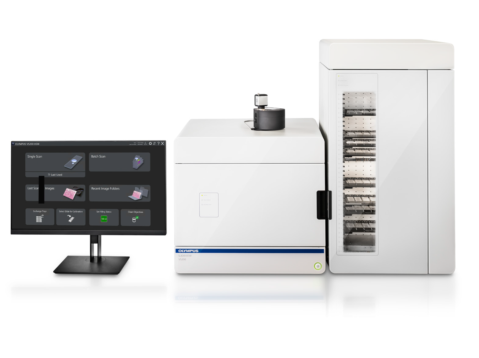
High-resolution whole-slide scanning and digital pathology support digital archiving, remote sharing and cloud-based analysis of H&E, IHC and multiplex fluorescence slides. AI-assisted image analysis and expert pathological interpretation can be combined to provide efficient, reliable data for research and drug development.
Quantitative Pathology
Image-analysis methods and AI algorithms support quantitative pathology from single-marker IHC to multiplex immunofluorescence. Objective, reproducible analysis of tissue features, immunolabeling and cellular phenotypes, together with batch processing and spatial analysis, helps turn pathology data into actionable research findings.

Research Pathology Reports
Qualified pathology specialists provide research pathology assessment and reports covering histological evaluation, toxicology, pharmacodynamic analysis and biomarker validation in preclinical studies. Digital pathology and quantitative image analysis can be incorporated to support research and drug development. Reports are for research use, not clinical diagnosis.
Tissue and Sample Submission Requirements
Follow the sample preparation and storage requirements below to support experimental quality and data reliability. Every sample must carry a clear label and a unique identifier. Before shipment, confirm that containers are securely sealed and labels are legible.
| Sample Type | Sample Requirements | Storage | Transport | Notes |
|---|---|---|---|---|
| Fresh tissue | Rinse with PBS to remove blood and debris, then immediately freeze at -80°C. | -80°C | Dry ice | Suitable for protein or RNA extraction. |
| Routinely fixed tissue | Use a fixative volume at least 10 times the tissue volume and preserve tissue morphology. Specify fixative type and fixation time. | Room temperature | Room temperature | Avoid compression or deformation. |
| Tissue requiring special fixation | Use the appropriate special fixative to preserve the required tissue structures. | Room temperature | Room temperature | Specify the fixative type where possible. |
| Paraffin tissue blocks | Use standard paraffin embedding cassettes with well-embedded tissue and no cracks. Blocks prepared within the past 6 months are recommended. | Room temperature | Room temperature | Older blocks may show uneven staining. |
| Tissue sections | Use adhesion-coated slides. Specify section thickness and slide-baking temperature. | 4°C | Ice packs | Suitable for immunohistochemistry and staining experiments. |
| Cells on coverslips | Culture cells on coverslips in 12- or 24-well plates and fix them. Wash 2–3 times with PBS and store sealed. | Room temperature | Room temperature | Ensure the plate-sealing film is tightly sealed. |
| Plant samples | Collect fresh material and fix in FAA. Fix leaf samples before sectioning. | Room temperature | Room temperature | Label the sampled part and specimen type. |
| Antibodies | Specify the antibody name and lot number and provide sufficient stock solution. | -20°C | Ice packs or dry ice | Avoid repeated freeze-thaw cycles. |