
Functional Evaluation of Cardiac Organoids and Cardiac Tissue-Like Models
Cardiac organoids and cardiac tissue-like preparations provide three-dimensional in vitro systems for studying cardiac function. We design assays around electrical activity, calcium signals and contraction to support model characterization, pharmacological response studies and functional mechanism research.
Cell composition, size and maturation vary with the construction method. Assay design considers tissue thickness, accessible regions and culture format to define what is measured and how broadly the results can be interpreted.
Functional Assay Capabilities
| Assay | Evaluation scope |
|---|---|
| Patch-clamp action potentials | Record membrane potential and analyze action potential duration, waveform and abnormal electrical activity. |
| Calcium imaging | Analyze calcium transient amplitude, kinetics and rhythm, together with spatial propagation in suitable preparations. |
| Voltage imaging | Observe optical electrical activity to assess repolarization, rhythm and spatial activity patterns. |
| Contractile function | Quantify contraction and relaxation, selecting motion-based or mechanical endpoints according to sample suitability. |
| Electrical-calcium-contraction coupling | Analyze electrical activity, calcium handling and contraction together to investigate where functional changes arise. |
These assay categories cover primary cardiomyocytes, iPSC-derived cardiomyocytes and cardiac organoid or cardiac tissue-like models when the preparation is suitable. The recording site, culture format and analysis parameters are chosen for the sample and study objectives; three-dimensional model compatibility is assessed for each method.
Assay Design for Three-Dimensional Models
| Assay component | Design considerations |
|---|---|
| Patch-clamp recording | Select accessible cells within the preparation or suitable dissociated cells according to recording access; distinguish cellular findings from conclusions about the entire tissue. |
| Calcium and voltage imaging | Define regions of interest with attention to probe penetration, optical access, motion and spatial sampling. |
| Contraction and coupling analysis | Select endpoints according to tissue anchoring and observable motion, then relate electrical activity, calcium handling and mechanical responses. |
Field-potential, impedance and flexible-substrate mechanical assays require additional compatibility assessment of tissue contact and culture morphology. Measurements obtained in one culture format are not assumed to apply to every three-dimensional model.
Calcium Signal Dynamics and Spatial Distribution
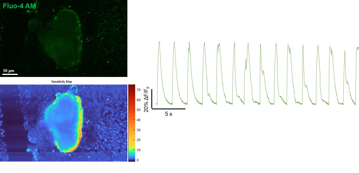
Calcium imaging can reveal local transients and regional differences in activity. Comparison with other functional endpoints can help investigate whether altered calcium handling is accompanied by electrical or contractile abnormalities.
Study Design and Deliverables
The model, treatment concentrations, exposure times, controls and replication are agreed according to the research objectives. Results are analyzed alongside sample condition and quality-control information.
- Records of model construction and sample characteristics, with descriptions of the measured regions and analysis methods.
- Representative images or signals and regional- or sample-level functional parameters.
- Interpretation that accounts for model heterogeneity and sampling coverage.